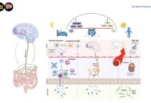
Holographic optogenetics could enable faster brain mapping for new discoveries
. Credit: Triplett et al. (Nature Neuroscience, 2025).")
Recent technological advances have opened new possibilities for neuroscience research, allowing researchers to map the brain’s structure and synaptic connectivity (i.e., the junctions via which neurons communicate with each other) with increasing precision.
Despite these developments, most widely employed methods to image synaptic connectivity are slow and fail to precisely record changes in the connections between neurons in vivo, or in other words, while animals are awake and engaging in specific activities.

Two different research groups, one based at Columbia University and UC Berkeley, and the other at the Vision Institute of Sorbonne University in Paris, introduced a promising approach to study synapses in vivo. Their proposed mapping strategies, outlined in two Nature Neuroscience papers, combine holographic optogenetics, a method to selectively and precisely stimulate or silence specific neuron populations, with computational techniques.
“This project was a really exciting collaboration between the Paninski and Adesnik labs at Columbia and UC Berkeley, aimed at developing much-needed tools for mapping how neurons in the brain are wired together,” Marcus A. Triplett, author of the first paper, told Medical Xpress.
“Understanding how the nervous system is wired up is important because that wiring is a large part of what gives the brain’s circuitry its function.”
Over the past few years, many neuroscientists have been trying to map large sections of brain tissue using a technique known as electron microscopy. This is a tool that utilizes applied beams of electrons to produce highly detailed images of biological samples, which can unveil significantly smaller brain structures than conventional light-based imaging methods.
“While these ventures have been highly successful in their own right, they provide a limited type of information because they map connections in fixed (non-living) tissue,” said Triplett.
“We wanted to develop a technique that had the potential to map large volumes, but that could also provide direct measurements of crucial biophysical variables, like the strengths of the connections between neurons, that are only available in living tissue.”
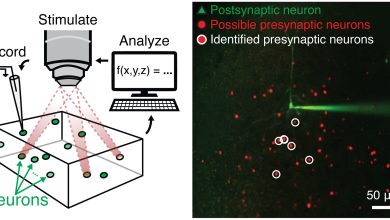
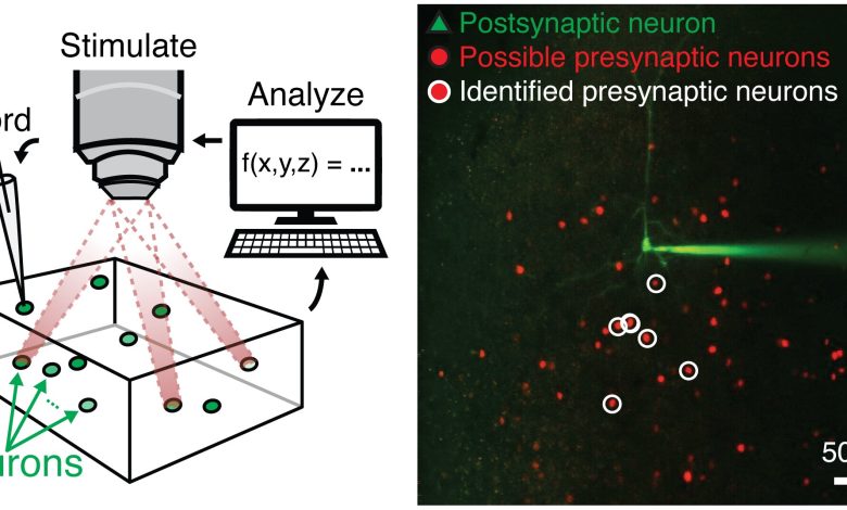
To probe for the existence of synaptic connections in brain slices, Triplett and his colleagues at Columbia and UC Berkeley used a technique known as holographic optogenetics, which uses light to activate specific sets of neurons where they introduced light-sensitive proteins called opsins.
By recording the electrical activity of one neuron while simulating another using optogenetic tools, neuroscientists can probe the connection between the two neurons. If two neurons are connected by a synapse, they would observe a transmission of neural activity, which essentially means that one neuron would elicit activity in the other.
“Our innovation in this study was to create a new computational method combining two important techniques from applied mathematics—namely, deep learning and compressed sensing,” explained Triplett. “These techniques enable the optogenetic approach to map connections between neurons an order of magnitude faster than previous approaches.”
Triplett and his colleagues assessed the potential of their holographic optogenetics technique in a series of tests and their results were very promising. In fact, they found that their approach could map 10 times the number of connections than those mapped by previously introduced approaches within the same timeframe.
“I think that our technique will see the greatest use in the context of studying neural computation—i.e., discovering how the brain’s wiring confers it with its remarkable computational abilities,” said Triplett. “Down the line, our method could also have important implications for understanding disease etiology, as neurological disorders can be associated with abnormal synaptic connectivity.”
Around the same time as Triplett and his colleagues, the “Wavefront Engineering Microscopy team” at Sorbonne University also started exploring the potential of holographic optogenetics as a tool to precisely control the activity of neurons and map the connections between them. Their work led to the introduction of various cutting-edge optical tools to study the brain’s structure and synaptic connectivity.
“Our team—combining expertise in physics, optical engineering, and neurobiology—has been among the pioneers in developing advanced optical methods to take fully advantage of these possibilities,” Dimitrii Tanese, co-author of the second paper, told Medical Xpress.
“We have introduced techniques to precisely shape light in space and time, allowing us to target and manipulate neuronal activity non-invasively within the living brain.”
.")
After testing the effectiveness of various optogenetic techniques for neuroscience research and applying them in their research, Tanese and his colleagues started looking for a new approach that would overcome their limitations. Specifically, they realized that existing techniques were ineffective for mapping connections between neurons in vivo and in real-time.
“Understanding how individual neurons are connected within a functional, living brain is often regarded as the holy grail of neuroscience, as it could reveal how network structure relates to function, how the brain reorganizes through plasticity, and how it recovers after injury,” said Tanese.
“We sought to exploit the possibilities opened by optogenetics, to overcome existing limitations and to establish and validate a scalable, quantitative framework for mapping synaptic connections directly in the intact brain with high precision and speed.”
To map connections between neurons, researchers need to be able to generate electrical signals and monitor how they propagate across synapses connecting pairs of neurons. Conventional approaches to doing this, which rely on the implantation of electrodes inside brain tissue, are both invasive and unable to probe several connections at once.
“Our goal is to exploit the low invasiveness, flexibility, and precision of light to accelerate this process,” explained Tanese.
“Specifically, we used an optical technology called two-photon holographic stimulation, which allows us to reshape light at will and precisely target specific cells of interest. Combined with the genetic expression of light-sensitive proteins (opsins), this technique enables us to ‘light up’ individual neurons deep inside the brain with pinpoint accuracy—like selectively pressing buttons in a three-dimensional circuit.”
While selectively activating a neuron inside the brain, the researchers recorded the electrical activity of another neuron, to determine whether it received a signal, which would in turn indicate the presence of a synaptic connection. As these connections are relatively rare, however, testing neurons one at a time is highly inefficient and time-consuming.
“To overcome this limitation, we explored the versatility of holographic light shaping to activate multiple neurons simultaneously, performing a form of group testing and, collaborating with Oweiss’s team (Univ. of Florida), we combined it with computational algorithms that reconstruct which neurons are truly connected,” said Tanese.
“Using our approach, we could map up to 100 presynaptic neurons in the intact mouse brain within five minutes and in a single experiment, representing order of magnitude improvement over previous approaches.”
Similarly to the technique employed by Triplett and his colleagues, the experimental approach devised by this research team draws from a wide range of fields, including neuroscience, electrophysiology, genetics, optics and signal analysis.
As part of their study, Tanese and his collaborators demonstrated the potential of their technique by using it to map synapses in the brain of live mice.
“By combining speed, precision, and scalability, our approach overcomes the main limitations of traditional electrophysiological techniques and opens new possibilities for studying how neural circuits are organized in the living brain,” said Tanese.
“In the long term, we believe this framework will help build a more integrated understanding of how neuronal networks support perception, adaptation, and cognition, bridging the gap between synaptic mechanisms, network structure and brain function.”
Overall, the new holographic optogenetics techniques developed by these two teams of researchers could soon open new exciting possibilities for neuroscience research. Tanese and his colleagues are now trying to improve their approach, for instance, by integrating the use of voltage indicators, fluorescent molecules that can pick up small changes in the electrical potential of neurons.
“Now that these sensors are becoming sensitive enough to detect signals as subtle as individual synaptic inputs, we will be able to eliminate the last remaining electrode from our experiments, achieving what can be defined as all-optical synaptic mapping,” added Tanese.
“In such a strategy, light would be used both to activate neurons and to monitor the resulting responses in their neighbors. This advancement would greatly reduce invasiveness while further increasing throughput, paving the way for large-scale circuit mapping and longitudinal studies that track connectivity changes over time during learning, experience, or disease progression.”
The other research team at Columbia and UC Berkeley are now also conducting further research aimed at improving their approach and using it to map synapses in specific brain regions.
“On the experimental side of things, we’re really excited about putting these tools to work to better understand the brain circuitry underlying visual perception,” said Triplett.
“On the computational side, we plan to keep scaling our techniques to map larger populations of neurons as quickly as possible. One cubic millimeter of the mouse brain contains tens to hundreds of thousands of neurons depending on brain region, and even one of those neurons can make thousands of synaptic connections across the brain, so there’s a lot of work to do!”
Written for you by our author Ingrid Fadelli, edited by Sadie Harley, and fact-checked and reviewed by Robert Egan—this article is the result of careful human work. We rely on readers like you to keep independent science journalism alive.
If this reporting matters to you,
please consider a donation (especially monthly).
You’ll get an ad-free account as a thank-you.
More information:
Marcus A. Triplett et al, Rapid learning of neural circuitry from holographic ensemble stimulation enabled by model-based compressed sensing, Nature Neuroscience (2025). DOI: 10.1038/s41593-025-02053-7.
I-Wen Chen et al, High-throughput synaptic connectivity mapping using in vivo two-photon holographic optogenetics and compressive sensing, Nature Neuroscience (2025). DOI: 10.1038/s41593-025-02024-y.
© 2025 Science X Network
Citation:
Holographic optogenetics could enable faster brain mapping for new discoveries (2025, November 3)
retrieved 3 November 2025
from https://medicalxpress.com/news/2025-10-holographic-optogenetics-enable-faster-brain.html
This document is subject to copyright. Apart from any fair dealing for the purpose of private study or research, no
part may be reproduced without the written permission. The content is provided for information purposes only.